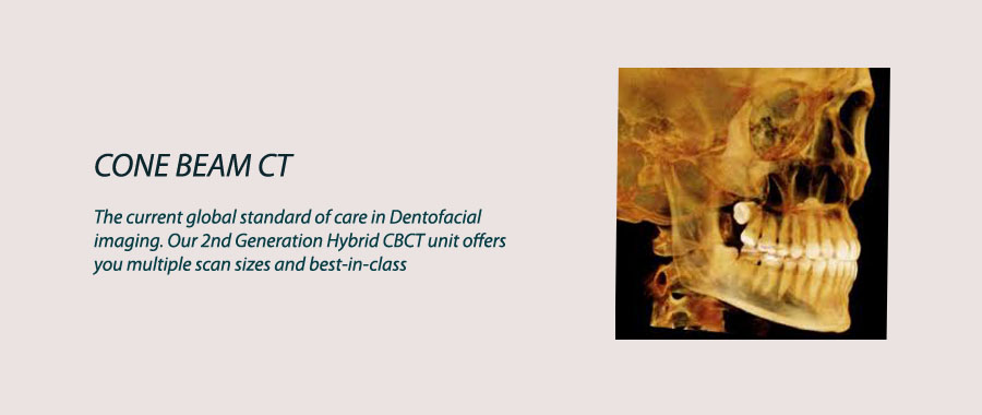
Cone Beam CT

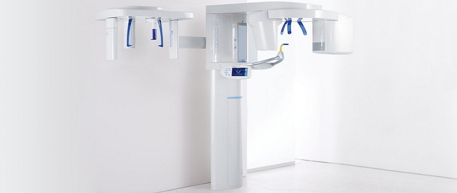
What is CBCT Scanning?
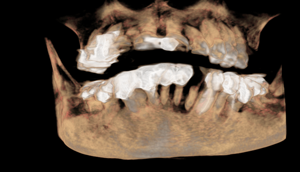
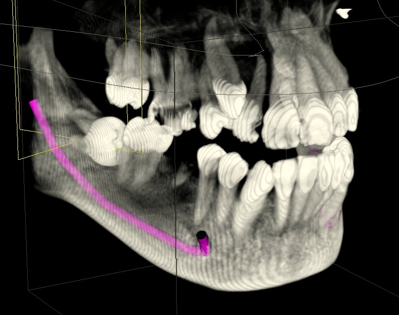
Cone beam CT provides high resolution, volumetric images that provide complete three-dimensional views of maxillofacial anatomy for more thorough analysis of bone structure and tooth orientation.
Cone beam CT delivers lower radiation dose, in-chair patient positioning (as opposed to a tunnel) and quicker scan times when compared to conventional medical CT Scan.
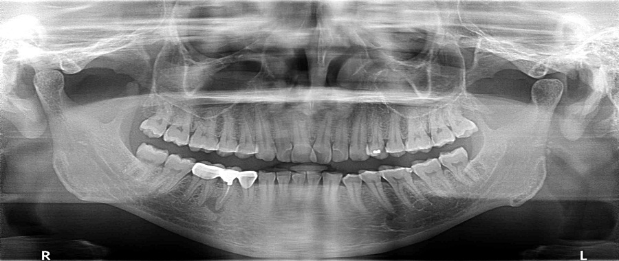
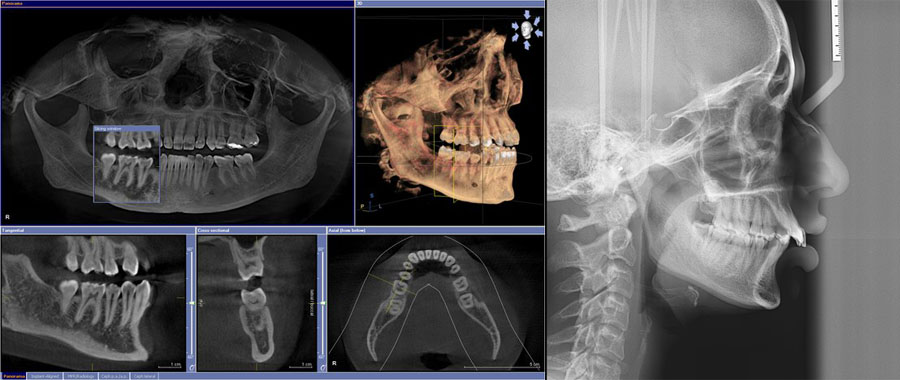
Why panoramic X-ray image is not enough?
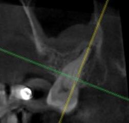
Panoramic images are magnified and distorted whereasCone beam images provide undistorted and accurate views of the jaws. As a result the use of panoramic images to perform measurements is unreliable. Another key difference is that CT images provide different views (cross-sectional, axial, coronal, sagittal, etc.) while panoramic x-rays provide only one view with all structures between the x-ray tube and the image detector superimposed on one another. This allows CT images to separate out the various structures.
What are the Main Applications of CT Imaging?
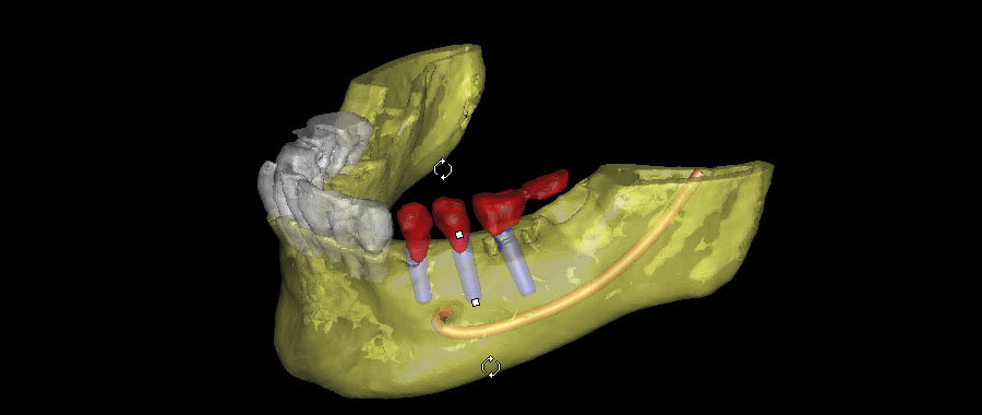
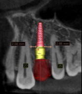
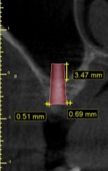
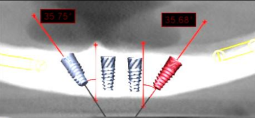
Dental Implants:
3D CT scans allow the surgeon and restorative dentist to optimally plan and place dental implants.
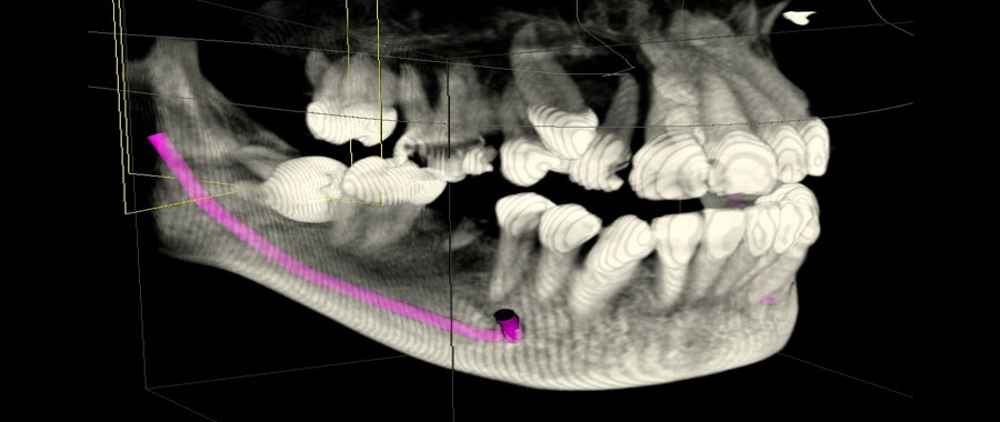
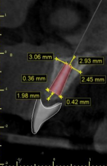
Pre-operative- identifying important landmarks like inferior dental nerve and maxillary sinus. To determine bone volume and density for optimum 3 dimensional placmenent.
Post operative- To assess bone loss and iatrogenic injury post implant plamcenent.
Periodontal:
To assess the prognosis of teeth in moderate to severe bone loss for comprehensive treatment planning.

Endodontics:
To assess complex root anatomy and root fractures.
Orthodontic:
Improve orthodontic diagnosis and treatment by providing the multiple projection perspective necessary to accurately assess tooth relationships.
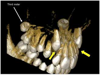
Impactions:
CBCT delivers precise 3D views of impacted molars within the alveolar bone, location relative to adjacent teeth, and proximity to vital structures, such as the nerve canal, sinus walls, and cortical borders.
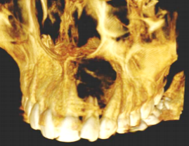
Pathology:
Assessing bone lesions and changes of the jaw, and detecting other pathologies, such as cysts, tumours, and disease.
Air Way assessment:
3D data enhances airway assessment and can result in reconsideration of the treatment plan if the patient has a typical airway, versus a restricted airway, which may be susceptible to collapse.
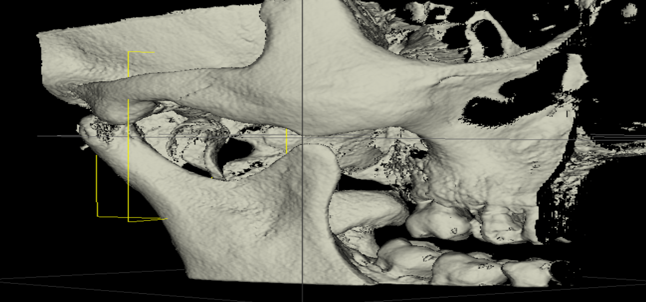
TMJ:
3D images allow the specialist to view critical structures for complete TMJ analysis and diagnosis clearly showing the condyles and surrounding structures, of bone morphology, joint space, and function all critical to TMJ dysfunction treatment and care.
Patient education:
We supply CT scan software free of charge with every CT scan. This software allows you to view, plan, measure, manipulate, print reports, create duplicates (for colleagues/patients) and, most important, show the actual treatment and assessments to your patients on your computer screen with all of his images and 3D images. The patient will understand the entire procedure and cannot fail to be impressed by the technology and your detailed assessment.
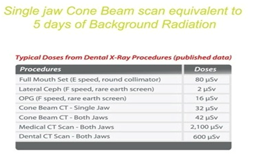
What is the radiation dose from Cone beam CT.